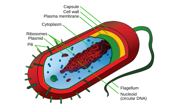
A typical bacterial cell – note the absence of a membrane-bound nucleus
This is the heart of Grade 10 Biology – the Cytology topic. LO 1 gave you the roadmap; now you're zooming in to study every room in the building. You'll compare plant, animal, protist, and bacterial cells at the organelle level, understand why structure determines function, and finish with a real-world case study on Tuberculosis (TB) – the disease that brings cell biology to life.
Essential Question: How does structure influence function?
Big Idea: Cells are the basic unit of structure and function in organisms. Every organelle has a specific structure engineered for a specific job – change the structure and you change (or destroy) the function.
Every living cell on Earth falls into one of two categories: prokaryotic or eukaryotic. The single most important distinction in all of cell biology.
A cell that lacks a membrane-bound nucleus and membrane-bound organelles. The DNA floats freely in the cytoplasm in a region called the nucleoid. From Greek: pro = before + karyon = kernel/nucleus. Examples: bacteria and archaea.
A cell that has a membrane-bound nucleus and membrane-bound organelles (mitochondria, ER, Golgi, etc.). From Greek: eu = true + karyon = kernel/nucleus. Examples: animals, plants, fungi, and protists.
Common exam trap: Not all unicellular organisms are prokaryotic! Amoeba, yeast, and many protists are unicellular eukaryotes – they HAVE a membrane-bound nucleus. Unicellular ≠ prokaryotic. And all prokaryotes are unicellular, but not all unicellular organisms are prokaryotes.
Despite the enormous differences between a bacterium and a human muscle cell, every living cell on Earth shares these four components:
The genetic material – the instruction manual for everything the cell does. In prokaryotes, DNA is circular and floats in the cytoplasm. In eukaryotes, DNA is linear and wrapped inside the nucleus.
The gel-like fluid filling the interior of the cell. All chemical reactions of life occur here. Contains water, salts, enzymes, and (in eukaryotes) organelles.
The selectively permeable barrier surrounding every cell. Controls what enters and exits. Made of a phospholipid bilayer with embedded proteins.
The protein-making machines. Found in ALL cells – prokaryotic and eukaryotic. Prokaryotes have smaller 70S ribosomes; eukaryotes have larger 80S ribosomes.
Exam favourite: "What do ALL cells have in common?" → DNA, cytoplasm, plasma membrane, and ribosomes. These four are universal – remove any one and the cell cannot survive.
You covered this in LO 1, but it's the foundation of everything here:
Observed dead cork cells under a microscope and coined the term "cell".
First to observe living cells – bacteria, protists, and sperm cells.
Concluded all plants are made of cells.
Concluded all animals are made of cells. Together with Schleiden, established cell theory.
Added the third principle: "Omnis cellula e cellula" – all cells come from pre-existing cells.
Bacteria are the simplest cells, but they're incredibly successful – they've been on Earth for over 3.5 billion years and live in virtually every environment.
A rigid outer layer surrounding the plasma membrane, made of peptidoglycan (a sugar-protein polymer unique to bacteria).
Many antibiotics work by attacking the bacterial cell wall. Since human cells lack cell walls, these antibiotics are selectively toxic – they kill bacteria without harming your cells. This is a key exam concept.
| Structure | Description |
|---|---|
| Nucleoid region | The area where circular DNA is concentrated – NOT enclosed by a membrane |
| Plasmids | Small circular DNA molecules separate from the main chromosome; carry genes for antibiotic resistance |
| Ribosomes (70S) | Smaller than eukaryotic ribosomes; site of protein synthesis |
| Cytoplasm | Gel-like fluid containing all cellular components |
| Cytoskeleton | Protein filaments that help maintain cell shape (simpler than eukaryotic cytoskeleton) |
Bacteria have NO membrane-bound organelles – no nucleus, no mitochondria, no ER, no Golgi, no chloroplasts, no lysosomes. This is the fundamental difference from eukaryotic cells. Though bacteria lack mitochondria, they perform aerobic respiration on the inner surface of their plasma membrane – this becomes critical when you study the Endosymbiotic Theory.
Eukaryotic cells are vastly more complex than prokaryotic cells. They are compartmentalised – different functions happen in different membrane-bound compartments (organelles), like departments in a company.


| Type | Examples | Unicellular or Multicellular | Cell Wall? |
|---|---|---|---|
| Animals | Humans, dogs, insects | Multicellular | No |
| Plants | Trees, grasses, ferns | Multicellular | Yes (cellulose) |
| Fungi | Mushrooms, yeast, moulds | Both | Yes (chitin) |
| Protists | Amoeba, Paramecium, algae | Mostly unicellular | Some do, some don't |
| Algae | Green algae, seaweed | Both | Yes (cellulose) |
The largest organelle in eukaryotic cells. Contains the cell's genetic material (DNA) organised into chromosomes. Enclosed by a double membrane called the nuclear envelope. Functions as the "brain" of the cell – controlling gene expression, growth, metabolism, and reproduction.

A double membrane (two phospholipid bilayers) surrounding the nucleus. Continuous with the rough endoplasmic reticulum (RER). Perforated by thousands of nuclear pores that regulate molecular traffic in and out of the nucleus.
Tiny channels in the nuclear envelope. Allow mRNA to exit (carrying instructions for protein synthesis) and allow proteins and ribosomes to enter. Think of them as security-controlled gates.
The combination of DNA + histone proteins inside the nucleus. Exists in two forms:
A dense region inside the nucleus (not membrane-bound). Responsible for producing ribosomal RNA (rRNA) and assembling ribosome subunits. The nucleolus is essentially a ribosome factory.
The gel-like substance inside the nucleus; equivalent to the cytoplasm but within the nuclear envelope.
The nucleus occupies about 10% of the cell's volume but controls 100% of its activities. Without the nucleus, the cell loses its ability to make proteins, repair itself, or divide – it dies. Red blood cells and platelets have no nucleus, which is why they are not considered true cells and have a limited lifespan.
A non-membranous organelle responsible for protein synthesis (translation). Made of rRNA and protein. Found in ALL cells – prokaryotic and eukaryotic.
| Feature | Prokaryotic Ribosomes | Eukaryotic Ribosomes |
|---|---|---|
| Size | 70S (smaller) | 80S (larger) |
| Location | Cytoplasm only | Cytoplasm, attached to RER, inside mitochondria (70S) & chloroplasts (70S) |
| Made by | Nucleoid region | Nucleolus |
Mitochondria and chloroplasts contain their own 70S ribosomes (the same size as bacterial ribosomes) – this is powerful evidence for the Endosymbiotic Theory (discussed later in this lesson).
An extensive network of membrane-enclosed channels extending from the nuclear envelope throughout the cytoplasm. The largest organelle by total membrane area. Comes in two types: Rough ER (RER) and Smooth ER (SER).
Called "rough" because its surface is studded with ribosomes.
Structure:
Functions:
Protein pathway: Ribosome on RER makes protein → protein enters RER lumen → protein is folded and partially glycosylated → packaged into a transport vesicle → sent to the Golgi apparatus for further processing.
A stack of flattened, membrane-bound sacs (cisternae) that receives, modifies, sorts, and ships proteins and lipids from the ER to their final destinations. Discovered by Italian physician Camillo Golgi in 1898.

Adds phosphate groups to proteins – this acts as an address label, directing the protein to its correct destination (plasma membrane, lysosome, or secretion outside the cell).
Adds or modifies carbohydrate (CHO) groups on proteins → produces glycoproteins. This is crucial for cell recognition and signalling.
Sorts modified proteins and lipids into vesicles for transport to:
Synthesises polysaccharides like cellulose (for plant cell walls) and other complex carbohydrates.
Determines the ratio of proteins to lipids in the plasma membrane. This is a critical regulatory function.
KEY EXAM CONCEPT: Which organelle controls the ratio/percentage of proteins vs lipids in the plasma membrane? It's the Golgi apparatus – NOT the RER and NOT the SER. The Golgi is the final decision-maker on what gets sent to the membrane.
Double-membraned organelles that generate most of the cell's ATP (adenosine triphosphate) through cellular respiration. Often called the "powerhouse of the cell." Contain their own DNA and ribosomes.

| Component | Description |
|---|---|
| Outer membrane | Smooth, permeable to small molecules |
| Inner membrane | Highly folded into cristae (singular: crista) – increases surface area for ATP production |
| Intermembrane space | Gap between outer and inner membranes |
| Matrix | The innermost compartment; contains enzymes for the Krebs cycle, mitochondrial DNA, and 70S ribosomes |
Mitochondria are described as an organelle that is "so so so rich" in features:
| Cell Type | Approx. Mitochondria Count | Reason |
|---|---|---|
| Muscle cell | ~1,000 | Extremely high energy demand for contraction |
| Liver cell | ~1,000–2,000 | High metabolic activity (detoxification, synthesis) |
| Lymphocyte (white blood cell) | ~5 | Relatively low energy demand |
| Red blood cell | 0 | No nucleus, no mitochondria – not a true cell |
The chemical equation for cellular respiration:
Glucose + Oxygen → Carbon Dioxide + Water + ATP (energy)
This is essentially the reverse of photosynthesis. Plants make glucose; mitochondria break it down to release energy.
A double-membraned organelle found in plant cells and algae that performs photosynthesis – converting solar energy into chemical energy (glucose/starch). Contains its own DNA and ribosomes. Like mitochondria, it is an autonomous "so so so rich" organelle.

| Component | Description |
|---|---|
| Outer membrane | Smooth outer boundary |
| Inner membrane | Encloses the stroma |
| Thylakoid | Flattened, disc-shaped membrane sacs where light reactions occur |
| Granum (plural: grana) | A stack of thylakoids – like a stack of coins |
| Stroma | The fluid-filled space between the inner membrane and thylakoids; site of the Calvin cycle |
| Own DNA | Circular, like bacterial and mitochondrial DNA |
| Own ribosomes (70S) | Like bacterial ribosomes – evidence for endosymbiosis |
| Starch granules | Storage of glucose as starch within the chloroplast |
Chloroplast converts solar energy → chemical energy (stored as food/starch). This is NOT called an "energy function" – the correct term is photosynthesis. The "energy organelle" (which converts chemical energy to ATP) is the mitochondrion.
Contains chlorophyll pigments (green). Performs photosynthesis to convert light energy into chemical energy (glucose/starch). Found in green parts of plants and green algae.
Contains carotenoid pigments (red, orange, yellow). Gives colour to flowers, fruits, and autumn leaves. Main function: attracting pollinators and seed dispersers through colour signals.
A non-pigmented (colourless) plastid used for storage – stores starch, lipids, or proteins. Found in roots, tubers, and seeds. Since it has no pigments, it is not involved in photosynthesis.
Exam tip: Chromoplast's function is attracting pollinators through colour (not photosynthesis). Leucoplast's function is storage (not photosynthesis). Only the chloroplast performs photosynthesis.
A membrane-bound organelle containing hydrolytic (digestive) enzymes capable of breaking down virtually any biological molecule. Functions as the cell's "digestive system" and "recycling centre." pH inside ≈ 2 (highly acidic). Present mainly in animal cells.
RER (makes the hydrolytic enzymes) → Golgi apparatus (packages them into vesicles) → Lysosomes (the finished product, ready for action)
| Type | Description |
|---|---|
| Primary lysosome | Newly formed, has not yet fused with anything; contains inactive enzymes |
| Secondary lysosome (Heterolysosome) | Formed when a primary lysosome fuses with a food vacuole or foreign particle (phagocytosis) |
| Autolysosome | Formed when a lysosome fuses with a damaged or worn-out organelle → recycles cellular components |
| Multivesicular body | Contains multiple smaller vesicles inside – involved in sorting and recycling |
Tay-Sachs Disease is a genetic disorder where lysosomes lack a specific lipase enzyme → lipids (gangliosides) accumulate in brain cells → progressive neurological damage → typically fatal in early childhood. This is a classic exam example of what happens when lysosomal function fails.
A single-membraned organelle that breaks down fatty acids and detoxifies harmful substances. Produces hydrogen peroxide (H₂O₂) as a by-product, then uses the enzyme catalase to convert it to water and oxygen. Abundant in liver and kidney cells.
Don't confuse peroxisomes with lysosomes! Lysosomes use hydrolytic enzymes at acidic pH to digest materials. Peroxisomes use oxidative enzymes to break down fatty acids and detoxify substances; their hallmark is producing and then neutralising H₂O₂.
The most prominent feature of a mature plant cell – can occupy up to 90% of the cell volume.
A dynamic network of protein filaments extending throughout the cytoplasm. Provides structural support, enables cell movement, facilitates intracellular transport, and plays a critical role in cell division.
| Type | Protein | Diameter | Functions |
|---|---|---|---|
| Microfilaments | Actin | ~7 nm (thinnest) | Cell shape, cytoplasmic streaming, cleavage furrow during cell division, muscle contraction |
| Intermediate filaments | Myosin and others | ~10 nm | Mechanical strength, anchoring organelles, resisting tension |
| Microtubules | Tubulin | ~25 nm (thickest) | Chromosomal movement during division (spindle fibres), intracellular transport, form cilia and flagella, form centrosomes |
Exam reminders:
A group of interconnected organelles that work together to synthesise, modify, package, and transport proteins and lipids. The system includes: nuclear envelope, endoplasmic reticulum (RER & SER), Golgi apparatus, lysosomes, vesicles/vacuoles, and the plasma membrane.
The starting point – continuous with the RER. Contains genes that code for proteins.
Synthesises proteins → folds them → packages them into transport vesicles → sends to Golgi.
Synthesises lipids and steroids → sends to Golgi for processing.
Receives products from ER → modifies (glycosylation, phosphorylation) → sorts → packages → ships to final destinations.
Receive digestive enzymes from the Golgi → carry out intracellular digestion and recycling.
Transport containers that shuttle materials between organelles and to/from the plasma membrane.
The final destination for many proteins and lipids. Composition controlled by the Golgi.
DNA (in nucleus) → Transcription → mRNA → exits through nuclear pore → Translation on ribosomes (attached to RER) → Polypeptide enters RER lumen → folded & partially glycosylated → Transport vesicle → Golgi apparatus (cis face) → modified & sorted → Golgi vesicle (trans face) → shipped to:
CRITICAL EXAM CONCEPT: An organelle is part of the endomembrane system based on its ORIGIN (where it came from physically), NOT its function. To be a member, the organelle must have originated from the ER, the Golgi, or be the nuclear envelope itself.
Mitochondria are NOT part of the endomembrane system – they originated from endosymbiosis (an engulfed bacterium), not from the ER/Golgi.
Chloroplasts are NOT part of the endomembrane system – same reason as mitochondria.
Ribosomes are NOT part of the endomembrane system – they originate from the nucleolus, not from the ER/Golgi.
The theory that mitochondria and chloroplasts were once free-living prokaryotes that were engulfed by a larger ancestral cell. Over time, a mutualistic relationship developed, and the engulfed prokaryotes became permanent organelles.

| Evidence | Explanation |
|---|---|
| Double membrane | The inner membrane = original prokaryote's membrane; outer membrane = the host's engulfing membrane |
| Own circular DNA | Like bacterial DNA (not linear like nuclear DNA) |
| Own 70S ribosomes | Same size as bacterial ribosomes (not 80S like other eukaryotic ribosomes) |
| Binary fission | Mitochondria and chloroplasts divide by binary fission – just like bacteria |
| Size | Similar in size to bacteria |
| Antibiotic sensitivity | Antibiotics that kill bacteria also affect mitochondria/chloroplasts (because of similar ribosomes) |
The same process explains chloroplasts: a large cell engulfed a photosynthetic cyanobacterium → the cyanobacterium became the chloroplast. This is why chloroplasts also have double membranes, own DNA, 70S ribosomes, and can self-replicate.
This is one of the most-tested topics in STEM Biology exams. Study this table carefully:
| Feature | Animal Cell | Plant Cell |
|---|---|---|
| Shape | Round/irregular | Rectangular/fixed (due to cell wall) |
| Cell wall | Absent | Present (made of cellulose) |
| Chloroplasts | Absent | Present (photosynthesis) |
| Central vacuole | Small/absent | Large (up to 90% of cell volume) |
| Centrosomes | Present | Generally absent |
| Lysosomes | Present and prominent | Rare (vacuoles serve similar function) |
| Plasma membrane | Present | Present (inside the cell wall) |
| Nucleus | Present | Present |
| Mitochondria | Present | Present |
| ER (RER & SER) | Present | Present |
| Golgi apparatus | Present | Present |
| Ribosomes | Present (80S) | Present (80S) |
| Cytoskeleton | Present (with centrosomes) | Present (without centrosomes) |
| Storage molecule | Glycogen | Starch granules |
| DNA location | Nucleus + Mitochondria | Nucleus + Mitochondria + Chloroplast |
Memory aids:
Cell wall present in:
Cell wall ABSENT in:
The cell wall provides rigidity and protection – this is why plant cells are rectangular and maintain their shape even when the cell membrane shrinks (plasmolysis). Animal cells, lacking a cell wall, are flexible and can change shape.
As a cell grows larger, its volume increases faster than its surface area. This means the cell membrane (surface) becomes insufficient to transport enough nutrients in and waste out for the increasing volume of cytoplasm. This is why cells stay small and organisms become multicellular instead of having one giant cell.
Imagine a cube with side length 1 cm:
Now a cube with side length 2 cm:
The SA:V ratio decreased – meaning less surface area per unit of volume. The larger cell would struggle to get enough nutrients and expel enough waste through its membrane. Solution? Divide into many smaller cells (→ multicellularity) or have highly folded membranes (→ cristae in mitochondria, microvilli in intestines).
Two organelles in eukaryotic cells are semi-autonomous – they can make some of their own proteins and replicate independently:
Radioactive labelling experiment: If you use radioactive thymine (which is incorporated only into DNA), you will find radioactivity in THREE locations in a plant cell: the nucleus, the mitochondria, and the chloroplasts – because all three contain DNA. In an animal cell, only TWO locations: the nucleus and the mitochondria.
| Category | Organelles |
|---|---|
| Double-membraned | Nucleus, Mitochondria, Chloroplasts |
| Single-membraned | ER (RER & SER), Golgi apparatus, Lysosomes, Peroxisomes, Vacuoles |
| Non-membranous | Ribosomes, Cytoskeleton (microfilaments, intermediate filaments, microtubules), Centrosomes |
TB is the real-world disease that connects cell biology to medicine. Everything you've learned about lysosomes, macrophages, and the immune system comes together here. TB is one of the oldest and most deadly diseases in human history – traces have been found in Egyptian mummies over 3,000 years old.
A chronic infectious disease caused by the bacterium Mycobacterium tuberculosis. Primarily attacks the lungs (pulmonary TB) but can spread to other organs (extrapulmonary TB). TB is the #1 infectious disease killer worldwide – killing approximately 3 million people per year.
The causative agent of TB. A rod-shaped bacterium (prokaryote) with an unusually thick, waxy cell wall containing mycolic acids – making it extremely resistant to drying, disinfectants, and many antibiotics. This waxy coat also prevents lysosomes from effectively killing it.
TB spreads through inhalation (airborne transmission) – breathing in tiny respiratory droplets containing the bacteria when an infected person coughs, sneezes, speaks, or sings.
This is different from malaria (which spreads via a vector – the mosquito). TB spreads directly from person to person through the air. One infected person in a crowded room can expose everyone nearby.

When you inhale Mycobacterium tuberculosis, your immune system responds by sending macrophages (a type of white blood cell) to the lungs to engulf and destroy the bacteria. What happens next depends on the strength of your immune system:
If your immune system is strong:
This is called a latent infection (عدوى كامنة) – the pathogen is present but "imprisoned." The person:
Think of it as a prison – the bacteria are alive but locked up. As long as the guards (immune system) stay strong, the prisoner stays contained. But if the guards weaken...
A person with latent TB can develop active TB later in life if their immune system weakens. The bacteria "escape" from the macrophages and begin multiplying.
Risk factors that convert latent TB → active TB:
| Risk Factor | Explanation |
|---|---|
| HIV/AIDS | Destroys the immune system. People with HIV are 50 times more likely to develop active TB |
| Diabetes mellitus | Weakens immune response |
| Silicosis | Occupational lung disease that damages lung tissue |
| Immunosuppressive drugs | Medications that deliberately weaken the immune system (e.g., after organ transplant) |
| Old age | Natural decline in immune function |
| Malnutrition | Weakened immune system due to lack of proper nutrition |
| Cancer | Both the disease and chemotherapy weaken immunity |
CRITICAL EXAM CONCEPT: An HIV-positive person is 50 times more likely to develop active TB than an HIV-negative person. AIDS = Acquired Immunodeficiency Syndrome – the word "deficiency" tells you the immune system is weakened. The risk of converting latent → active TB is highest in the first 2 years after initial infection, then gradually decreases (but never reaches zero).
| Term | Meaning |
|---|---|
| Primary TB | The initial infection – may be latent (contained) or active (immediate disease) |
| Post-primary TB (Secondary/Reactivation TB) | When latent TB reactivates into active TB – the bacteria escape from macrophages |
| Cavitary TB | Active TB that causes cavities (holes) in the lung tissue where bacteria escaped from macrophages – visible on chest X-ray |
| Fibro-cavitary lung lesion | The characteristic X-ray finding of post-primary TB – cavities surrounded by fibrous tissue |
Cavitation (formation of cavities in the lungs) is the hallmark of post-primary TB (reactivation from latent to active). It occurs at the site where bacteria escape from macrophages. If an exam asks: "Fibro-cavitary lesion on X-ray is a feature of ___?" → The answer is post-primary (secondary) TB, NOT primary TB.
Latent infection CAN become active infection (if immunity drops). But active infection CANNOT revert back to latent infection – once the bacteria are free and multiplying, they can't be "re-imprisoned." Treatment with antibiotics is required.
Pathway: TB Bacteria → Infection → either Latent (no disease) or Active (TB disease)
| Method | Details |
|---|---|
| Tuberculin skin test (TST) | Injection of tuberculin protein under the skin. If hypersensitivity (swelling) develops → positive → person has TB bacteria. But this test cannot distinguish between latent and active TB. |
| Chest X-ray | Can reveal cavitary lesions (cavities in the lungs) characteristic of active or post-primary TB |
| Sputum microscopy | Direct examination of sputum (phlegm) under a microscope to find TB bacteria |
Since TB is caused by a bacterium, it is treated with antibiotics. Two key antibiotics:
| Antibiotic | Mechanism |
|---|---|
| Isoniazid (أيزونيازيد) | Interferes with the bacteria's ability to make their cell wall → bacteria cannot maintain structural integrity → die |
| Rifampicin (ريفامبيسين) | Prevents the bacteria from making proteins (inhibits RNA polymerase) → bacteria cannot function → die |
Why can't you use an antibiotic that "disrupts the nuclear envelope"? Because TB is caused by a bacterium – a prokaryotic cell that has no nuclear envelope! Any antibiotic targeting the nuclear envelope would be useless against bacteria. Exam writers love to test this connection between cell biology and medicine.
| Feature | Tuberculosis | Malaria |
|---|---|---|
| Causative agent | Mycobacterium tuberculosis (bacterium) | Plasmodium species (protozoan parasite) |
| Mode of transmission | Inhalation (airborne) | Vector (female Anopheles mosquito bite) or blood transfusion |
| Target organ | Lungs (primarily) | Liver (incubation) → Red blood cells (disease) |
| Type of infection | Can be latent or active | Always active (no latent form) |
| Treatment | Antibiotics (isoniazid, rifampicin) | Antimalarial drugs (quinine, chloroquine, artemisinin) |
| Classification | Endemic infectious disease | Endemic infectious disease |
| Resistance problem | Antibiotic-resistant TB (MDR-TB) | DDT-resistant mosquitoes |
This is a fascinating connection between genetics and infectious disease that ties back to cell structure.
A genetic disease where red blood cells take on an abnormal sickle (crescent) shape instead of the normal biconcave disc shape, due to a mutation in the haemoglobin protein.
People with sickle cell anaemia have natural resistance to malaria. Why?
This is a remarkable example of how one disease (sickle cell) can provide protection against another disease (malaria) – and how cell structure (the shape of RBCs) directly affects cell function (whether a parasite can invade).
Questions sourced from past STEM exams, reference materials, and medical question banks. Use these to test your understanding of everything covered in this lesson – cell structure, organelle functions, plant vs animal cells, endomembrane system, endosymbiosis, and TB.
Cell Structure & Organelles:
Plant vs Animal Cells:
Endomembrane System & Endosymbiosis:
Tuberculosis Case Study:
Video Resources: