This is your foundation for everything in Biology. Think of this lesson as the master mind map – it introduces cells, disease, microscopes, and the real-world case studies (Malaria & Sickle Cell) that tie the entire unit together. Every single LO in this unit builds on what you learn here. Nail this, and the rest falls into place.
The scientific study of living organisms – their structures, functions, interactions with each other, and relationships with their environment. From the Greek bios (βίος) = life + logos (λόγος) = study.
Biology covers everything from the tiniest bacteria to the largest whale. You'll study how organisms are built, how their systems work, how they interact with their environment, and how changes – like disease – disrupt everything.
Biology teaches you two critical skills: patience (especially if you're coming from an Arabic-language background – the terminology takes time) and information organisation. Biology isn't a "memorisation subject" or a "comprehension subject" – it's an information organisation subject. The student who organises their knowledge best scores highest.
Before we dive into disease, you need a mental map of how your body is organised – because everything in Biology traces back to one place: the gene.
The whole organism – you. Your body is a cooperative of trillions of specialised cells working together.
Your body is made of organ systems – the digestive system, nervous system, immune system, cardiovascular system, and more. Each system performs a major life function.
Each system contains organs – the stomach and pancreas in the digestive system, the heart and blood vessels in the cardiovascular system.
Organs are made of tissues – groups of similar cells working together to perform a specific function.
Tissues are made of cells – the fundamental unit of life. This is the smallest level that is still "alive."
Inside each cell, the nucleus (النواة) contains chromosomes. Each chromosome is made of DNA wrapped around proteins called histones (like thread wound around a spool). A gene is a specific segment of that DNA – responsible for a particular trait.
Your DNA is about 2 metres long, yet it fits inside a cell that's only a few micrometres wide. How? The DNA wraps around histone proteins – just like thread wrapping around a spool – allowing it to pack tightly into chromosomes.
A gene's function is called protein synthesis (تخليق البروتين):
This sequence – Gene → Transcription → mRNA → Translation → Protein → Trait – is the central pathway of molecular biology. You'll study it in detail in LO 5 (Protein Synthesis), so don't worry about memorising the mechanism now. Just understand the big picture.
The basic unit of structure and function in all living organisms. All organisms are made of one or more cells, and all cells arise from pre-existing cells.
Every cell has two main structural components:
You'll study cell structure in massive detail in the Cytology topic (LO 2–3) – the most important topic in Grade 10 Biology. What you're seeing here is just the roadmap.
English scientist Robert Hooke used a microscope to examine a thin slice of cork (tree bark). He observed tiny box-like chambers and called them "cells" because they reminded him of the small rooms (cellae) where monks lived. These were actually the dead cell walls of plant cells. Hooke is considered the first person to observe and name cells.
Dutch scientist Antonie van Leeuwenhoek built one of the first powerful microscopes and discovered bacteria (the smallest living organisms), single-celled protists, and sperm cells. He called the tiny organisms he saw "animalcules" (little animals).
German physiologist Theodor Schwann demonstrated that all animals are made up of cells, establishing the cellular basis of animal life.
German botanist Matthias Schleiden concluded that all plants are made up of cells, establishing the cellular basis of plant life.
Every living thing – from bacteria to blue whales – is built from cells. The cell is where life happens.
Unicellular organisms (like bacteria and amoeba) consist of a single cell. Multicellular organisms (like plants and animals) contain billions of specialised cells working together.
New cells don't appear from nothing – they come from the division of existing cells. This is how organisms grow, repair damage, and reproduce.
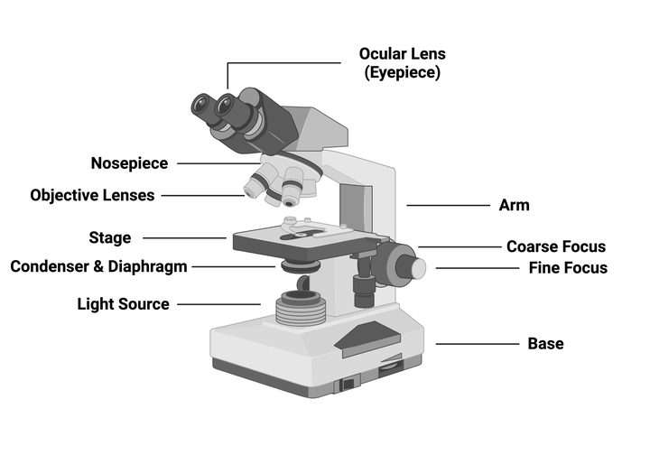
An instrument used to magnify objects too small to see with the naked eye. Microscopes are the primary tool biologists use to study cells and microorganisms.
Without microscopes, cell theory would never have been developed, and we would know nothing about the cellular basis of disease.
Uses visible light and glass/plastic lenses to magnify specimens.
This is the type Hooke and van Leeuwenhoek used – and the type you will use in your lab investigations.
| Property | Meaning | Why It Matters |
|---|---|---|
| Magnification (التكبير) | The ratio between the image size and the real size of the specimen | Higher magnification = larger image = more detail visible |
| Contrast (التباين) | The difference in brightness and darkness in the image | Better contrast = easier to distinguish different structures |
| Resolution (الدقة) | The ability to distinguish two close points as separate | Higher resolution = sharper, clearer image (not just bigger!) |
Exam trap: Magnification and resolution are NOT the same thing! You can magnify an image 10,000× but if the resolution is low, you'll just get a bigger blur. Resolution is what gives you clarity.
A disturbance, breakdown, or abnormality in the structure or function of an organism (or any part of it) that impairs normal health. In simpler terms – something goes wrong in the body that makes it not work properly.
All diseases fall into one of two categories:
How to tell them apart: Look at the cause. If a pathogen (bacteria, virus, fungus, parasite) is involved → infectious. If the cause is genetics, environment, lifestyle, or age → non-infectious. This is the #1 distinguishing factor – not whether the person is near others.
Diseases that cannot be transmitted from one person to another. They are NOT caused by pathogens but by factors such as genetics, malnutrition, environment, and lifestyle.
Exposure to pollution, toxic chemicals, or radiation can cause non-infectious diseases.
Examples:
Exam question (2024): Students were shown an image of factories emitting smoke and asked whether the resulting disease would be infectious or non-infectious. Answer: Non-infectious – because pollution is an environmental factor, not a pathogen.
| Disease | Arabic | Description |
|---|---|---|
| Diabetes (السكري) | مرض السكر | Body cannot properly regulate blood sugar due to insulin problems |
| Cancer (السرطان) | السرطان | Uncontrolled cell growth forming tumours that invade healthy tissue |
| Cardiovascular Disease | أمراض القلب والأوعية الدموية | Diseases of the heart and blood vessels (cardio = heart, vascular = blood) |
| Chronic Respiratory Disease | أمراض التنفس المزمنة | Long-term diseases of the lungs (respiratory = breathing, chronic = long-lasting) |
Diseases caused by pathogenic micro-organisms (bacteria, viruses, fungi, or parasites) that can be transmitted from one person to another. Also called communicable diseases.
A micro-organism that causes disease. The word comes from Greek: pathos (πάθος) = suffering + gen = producing. Literally: "something that produces suffering."
The smallest pathogens – they're not even technically "alive" because they can't reproduce on their own.
Examples of viral diseases:
The method by which a pathogen reaches and enters a new host. Understanding modes of infection is key to preventing disease transmission.
Breathing in pathogens suspended in the air – especially from an infected person's coughs or sneezes. Respiratory droplets can carry millions of pathogens.
Example: Tuberculosis, COVID-19, influenza
Swallowing contaminated food or water containing pathogens.
Example: Cholera (contaminated water), Salmonella (contaminated food)
Physical contact with an infected person or contaminated surface – touching, handshaking, or sharing personal items.
Example: Worm infections (pinworms spread by hand contact), skin infections
Transmission through intimate physical contact and exchange of bodily fluids.
Example: HIV/AIDS, sexually transmitted infections (STIs)
Accidental transmission during medical procedures – contaminated needles, blood transfusions, or organ transplants.
Examples:
A vector is a living organism (usually an arthropod like a mosquito or fly) that carries and transmits a pathogen from one host to another.
Example: The female Anopheles mosquito transmits malaria – the most important vector-borne disease in this LO.
The word "vector" comes from Latin vehere = "to carry." Think of vectors as transport vehicles for pathogens.
Critical distinction: Vectors do NOT cause disease – they only transport the pathogen! The pathogen (e.g., Plasmodium) causes the disease. The vector (e.g., mosquito) is just the delivery vehicle. This is a common exam trap.
A model showing the three essential components needed for most infectious diseases to spread: the pathogen, the vector, and the host.
The triangle works like this:
The disease-causing organism – a bacterium, virus, fungus, or parasite that initiates the infection.
The carrier organism (usually an insect) that picks up the pathogen and delivers it to the host. Not all infections use vectors, but many of the most dangerous ones do (malaria, dengue, Zika).
The organism that harbours the pathogen and provides it with shelter and nourishment. The host is where the disease actually manifests.
How it works: The pathogen lives in the environment or in a reservoir → a vector picks it up → the vector bites/contacts the host → the pathogen enters the host → infection occurs.
Breaking the triangle = stopping the disease. If you can eliminate ANY one side of the triangle – kill the pathogen, remove the vector, or protect the host – the chain of infection is broken. This is the basis of all disease control strategies.
The degree of damage a pathogen can cause to its host. Measured by the toxins it produces and its ability to invade tissues. Higher virulence = more dangerous pathogen.
Both true pathogens and opportunistic pathogens can cause infection – just under different conditions. A true pathogen attacks healthy people; an opportunistic pathogen waits until defences are down. Don't fall for the trap of thinking only one type causes disease!
The community of micro-organisms that naturally live in and on the human body without causing disease under normal conditions. They are part of the body's natural environment and many are actually beneficial.
Example: E. coli bacteria in your small intestine – they produce Vitamin K that your body needs, while you provide them with shelter and food. This is a mutually beneficial relationship (mutualism) – until your immune system weakens, and then the E. coli can become an opportunistic pathogen.
A living organism – most commonly an arthropod (like mosquitoes, flies, or ticks) – that transmits an infective pathogen from one host to another. From Latin vehere = "to carry."
Think of vectors as transport trucks. They pick up the pathogen from one location and deliver it to another. The truck doesn't create the cargo – it just moves it. Similarly, vectors don't cause disease – they only carry the pathogen.
An organism that harbours a pathogen and provides it with shelter (مأوى) and nourishment (تغذية). Any organism carrying a pathogen can be called a host.
Another way to put it: any organism that carries a pathogen = host.
The type of host depends on what changes happen to the parasite inside it:
The host inside which the parasite undergoes sexual reproduction.
Key characteristic: The parasite mates (sexual reproduction) inside this host.
Malaria example: The female Anopheles mosquito is the definitive host – Plasmodium parasites undergo sexual reproduction inside the mosquito's stomach.
Yes, the mosquito is the definitive host for malaria – not the human. This surprises many students! The defining criterion is where sexual reproduction occurs.
How to classify a host – the decision tree:
Yes! Since vectors carry pathogens, they are technically hosts too. A vector can be classified as either:
But a vector CANNOT be a reservoir host or transport host (because in vectors, the parasite always develops/reproduces).
A close, long-term relationship between two different organisms living together. The nature of the relationship depends on whether each organism benefits, is harmed, or is unaffected.
Both organisms benefit from the relationship.
The "T" in mutualism can help you remember: Together – both benefit together!
Example: E. coli bacteria in the human small intestine:
Both sides win = Mutualism ✅
| Relationship | Parasite/Organism A | Host/Organism B |
|---|---|---|
| Mutualism | ✅ Benefits | ✅ Benefits |
| Commensalism | ✅ Benefits | ➖ Unaffected |
| Parasitism | ✅ Benefits | ❌ Harmed |
Notice that the parasite always benefits in all three relationships – that's why it chooses to live on/in the host in the first place. The difference is what happens to the host.
For an infection to occur, three conditions must be met:
The micro-organism must successfully get inside the target tissue or organ – whether it's the liver, blood, lungs, or gut.
The body's immune system (white blood cells, antibodies) will try to destroy the invader. The pathogen must evade or overcome these defences to survive.
A single pathogen alone usually can't cause disease. It must reproduce and increase in numbers to establish an infection and cause symptoms.
Exam trap: A question may ask if the pathogen "must be a true pathogen" as a requirement. No! Both true AND opportunistic pathogens can cause infection – just under different conditions. Virulence type is NOT a requirement for infection.
A combination of multiple signs AND symptoms that regularly occur together and characterise a particular disease. Example: Down Syndrome involves a specific set of physical and developmental features.
A disease constantly present in a population or region at a low, steady rate.
A sudden increase in disease cases concentrated in a specific area (localised outbreak).
An epidemic that has spread across multiple countries or continents, affecting a massive number of people worldwide. Example: COVID-19 after it spread globally – it went from epidemic to pandemic.
| Type | Source | Example |
|---|---|---|
| Exogenous (خارجي) | Pathogen comes from outside the body – another person, animal, or environment | Salmonella from contaminated food |
| Endogenous (داخلي) | Pathogen already exists inside the body as part of normal flora but becomes pathogenic when immunity drops | E. coli UTI when immune system weakens |
| Zoonotic (حيواني المصدر) | Pathogen transmitted from animals to humans (zoo = animal) | Rabies (داء الكلب) from dog bites |
Rabies (السعار) is one of the most terrifying zoonotic infections. After a bite from a rabid animal, the virus attacks the nervous system, causing hydrophobia (fear of water – hydro = water, phobia = fear). The infected person becomes unable to swallow water, leading to death. There is no cure once symptoms appear – only prevention through vaccination.
Think: Like a storm – intense but brief.
The infection stays restricted to one area – it doesn't spread beyond the initial site.
Example: An eye infection that stays in the eye only.
Transmission between individuals in a population – from person to person, regardless of family relationship.
Transmission from parent to offspring – through genetics, birth, or breastfeeding.
Exam favourite! Most of your body has bacteria living in or on it (normal flora). But certain organs/sites are completely microbe-free under healthy conditions:
If pathogens reach any of these sites, it's a serious medical emergency.
When beneficial micro-organisms already living in the body prevent harmful pathogens from establishing infection. The "good" microbes compete with and block the "bad" ones – like a nightclub bouncer keeping troublemakers out.
An inanimate (non-living) object that can carry and transmit pathogens from one person to another. Examples: doorknobs, pens, phones, utensils, shared towels. A fomite is NOT a host – it's not alive.
Sickle Cell Disease is a non-infectious, genetic disease – it cannot be caught from another person. It is inherited from parents to children through a recessive gene mutation.
An inherited genetic disorder caused by a mutation in the beta-globin gene that changes the structure of haemoglobin protein in red blood cells. The abnormal haemoglobin causes RBCs to become rigid and form a sickle (banana/crescent) shape instead of the normal disc shape.

A mutation in the beta-globin gene (located on chromosome 11) produces abnormal haemoglobin called HbS instead of normal HbA.
The HbS proteins stack on top of each other inside the red blood cell, forming rigid chains.
The stacked proteins force the normally round, flexible RBC into a stiff, sickle/banana shape.
Sickle cells are fragile (break easily → anaemia), sticky (clump together → block blood vessels), and rigid (can't squeeze through narrow capillaries → pain and organ damage).
Sickle Cell Disease is caused by an autosomal recessive gene – meaning a person must inherit two copies of the mutated gene (one from each parent) to develop the full disease.
The Malaria-Sickle Cell Connection: People with sickle cell trait (one copy of the gene) have significant protection against malaria. The Plasmodium parasite has difficulty surviving inside sickle-shaped RBCs. This is why the sickle cell gene is most common in regions where malaria is prevalent (Sub-Saharan Africa) – it provides a survival advantage. This is a powerful example of natural selection.
The spleen is an organ that filters blood and fights infection. In sickle cell disease, sickle cells damage the spleen, making patients highly vulnerable to severe infections. The leading cause of death in children with sickle cell disease is pneumonia (not malaria) – because their damaged spleen can't fight bacterial infections.
Malaria is an infectious, vector-borne disease – it spreads through the bite of infected mosquitoes. It is one of the deadliest diseases in human history, killing hundreds of thousands of people every year, mostly children in Sub-Saharan Africa.
A serious, life-threatening infectious disease caused by Plasmodium parasites, transmitted through the bite of an infected female Anopheles mosquito. The parasites infect and destroy red blood cells, causing fever, anaemia, and potentially death.
An infected female Anopheles mosquito bites a human. The mosquito's saliva contains Plasmodium sporozoites (the infective stage), which enter the human bloodstream.
Through the mosquito bite, Plasmodium sporozoites enter the human blood and travel to the liver.
Sporozoites invade liver cells and undergo asexual reproduction, multiplying into thousands of merozoites. This is the incubation period – the patient feels nothing yet.
Merozoites burst out of liver cells and invade red blood cells (RBCs), where they feed on haemoglobin, multiply, and destroy the RBCs. This cycle of invasion → multiplication → destruction repeats, releasing more merozoites to infect more RBCs. This is when symptoms appear.
Some merozoites develop into gametocytes (male and female sexual forms) inside the human blood.
When another mosquito bites the infected human, it ingests the gametocytes. Inside the mosquito's stomach, the male and female gametocytes undergo sexual reproduction (sporogony), producing new sporozoites that migrate to the mosquito's salivary glands – ready to infect the next person.
| Organism | Host Type | Why? |
|---|---|---|
| Human | Intermediate host | Plasmodium undergoes only asexual reproduction in humans (liver and blood stages) |
| Female Anopheles mosquito | Definitive host AND vector | Plasmodium undergoes sexual reproduction (sporogony) in the mosquito's stomach |
Symptoms usually start 10–15 days after the mosquito bite (but can take weeks or months for some species).
The Classic Malaria Attack (Paroxysm):
The patient experiences intense, uncontrollable shivering and chills.
Body temperature spikes to dangerously high levels (40°C+).
Profuse sweating occurs, and the fever drops. The cycle then repeats.
Other symptoms: Headache, muscle pain, fatigue, nausea, vomiting, diarrhoea, abdominal pain, jaundice (yellow skin/eyes), anaemia, and in severe cases – confusion, seizures, and coma (cerebral malaria).
Some Plasmodium species (P. vivax & P. ovale) can stay dormant in liver cells for months to over a year – then symptoms suddenly appear long after the initial bite. This is why malaria diagnosis can be tricky.
| Method | Description | Speed |
|---|---|---|
| Microscopic examination (blood smears) | Gold standard – thick and thin blood smears viewed under a microscope show parasites inside RBCs | Moderate |
| Rapid Diagnostic Tests (RDTs) | Detect parasite antigens in blood | Results in < 15 minutes |
| PCR (molecular test) | Very accurate DNA-based test – identifies species and drug resistance | Slower, more expensive |
Treatment depends on the Plasmodium species, severity, patient's age, pregnancy status, and drug resistance in the area.
| Drug | Use |
|---|---|
| ACTs (Artemisinin-based Combination Therapies) | First-line treatment for P. falciparum (e.g., artemether-lumefantrine) |
| Chloroquine | For P. vivax, P. ovale, P. malariae (resistance common in P. falciparum) |
| Quinine sulfate | Used in severe/complicated cases |
| Atovaquone-proguanil (Malarone) | Good for both prevention and treatment |
Drug resistance is a major global problem, especially chloroquine resistance in P. falciparum. Treatment must be completed fully to prevent relapse and the development of further resistance.
Sleeping under treated nets prevents mosquito bites during the night (when Anopheles mosquitoes are most active).
Spraying interior walls of houses with insecticides kills mosquitoes that rest on walls.
Taking preventive anti-malarial medicines when travelling to malaria-endemic areas.
Wearing long clothes, using mosquito repellent, and avoiding outdoor activity at dawn/dusk.
Draining stagnant water where mosquitoes lay their eggs – puddles, old tyres, open containers.
This is a two-way street: poverty causes more disease, and disease makes poverty worse. This creates a vicious cycle that's extremely hard to break.
Countries with low income / developing countries face:
| Direction | Result |
|---|---|
| Poor economy → Disease | Bad water, no sanitation, no vaccines, malnutrition, weak health system → infectious diseases thrive |
| Disease → Poor economy | Fewer workers, high medical costs, less investment, less tourism, brain drain → economy weakens |
| Breaking the cycle | Economic improvement + strong public health investment → infectious diseases drop → but chronic diseases may rise later (the "developed country" pattern) |
Questions sourced from past STEM exams, reference materials, and medical question banks. Use these to test your understanding of everything covered in this lesson.
Cell Theory:
Microscopes and Their Types:
Infectious and Non-Infectious Diseases:
Malaria Case Study:
Sickle Cell Disease:
Additional Video Resources:
External Reference: